

Arthroscopic surgery, also known as “keyhole” surgery, is a type of minimally invasive surgery (MIS) often offered by orthopaedic surgeons to patients who are suffering from a variety of joint injuries and degenerative conditions. By using a tiny high-resolution camera to look inside the joints, the surgeon is able to diagnose and treat many joint conditions.
In this article, we’ll be sharing about how arthroscopic surgery is performed, what conditions can be treated with this surgery, as well as its benefits and risks.
Prior to arthroscopic surgery, your doctor will examine the affected joint thoroughly, and ask you some questions about your condition. Additional imaging may be performed, such as an X-ray or MRI.
As its name suggests, “keyhole” surgery involves a tiny slit-shaped incision being made to access the affected joint, which is usually less than a centimetre. A tiny camera is inserted through the incision to look inside the joint and allow the surgeon to visually inspect the site of injury or disease under magnification.
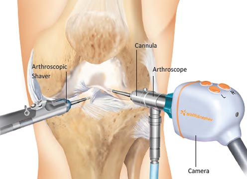
Once the diagnosis has been confirmed, an additional sub-centimetre “keyhole” incision may be made to insert specialised instruments, which the surgeon will use to treat the problem. In the hands of a trained arthroscopic surgeon, this “keyhole” technique allows for accurate diagnoses to be made, and facilitates the repair and reconstruction of many joint injuries and diseases.
After the operation, you can expect the small wounds to take approximately a week to heal. In addition to barely visible scars, arthroscopic surgery has the advantages of little pain and rapid recovery of joint function owing to the minimal injury to skin and soft tissues during the operation.
As part of accelerated recovery post-operatively, physiotherapy and occupational therapy may be offered to you as well, to help you regain strength, stability and full range of motion of the joint more quickly.
- Less invasive & lower chance of infection – This minimally-invasive surgical (MIS) procedure only leaves small slit-like wounds (less than 1cm), substantially lowering the risk of wound complications and infections.
- Less pain & swelling – Less soft tissue is damaged during this minimally-invasive surgery (MIS), substantially reducing the amount of post-operative pain, inflammation, swelling and stiffness.
- Faster recovery time – Compared to open surgery, arthroscopic / “keyhole” surgery a significantly shorter recovery time and will allow you to resume normal activities almost immediately.
- Better cosmesis – In open surgery, a large & unsightly scar often remains; however, in “keyhole” surgery, the wounds are small (less than 1cm), slit-like and usually barely visible, achieving far better cosmetic outcomes.
- Selected indications – Although “keyhole” surgery is suitable for most patients, severe joint deformities or multiple previous surgeries can limit joint access and arthoscopic surgery may not be suitable in this small group of patients.
- Limited availability – Compared to conventional open surgery, arthroscopic “keyhole” surgery is more technically demanding, which requires a highly trained and skilled surgeon to perform.
Arthroscopic “keyhole” surgery is a very safe procedure (MIS) and has a low risk of side effects and complications. Nevertheless, as with any surgery, there are some rare risks that patients should be aware of:
- Infection – The wound is small, but there is still a chance of infection, although it is far lower than in open surgery.
- Swelling of the joint – Haemarthrosis, or bleeding into the joint, may occur shortly after the operation, and cause discomfort and some swelling; this risk is lower than in open surgery owing to the minimally-invasive nature of “keyhole” surgery
- Stiffness – Some patients may have residual joint stiffness after surgery, but this is less likely and less severe than after open surgery, as there is much less soft tissue injury in arthroscopic surgery.
- Injury to surrounding muscles and nerves – This may lead to numbness or pain, but the risk of this occurring is low, as the incisions are small and carefully planned to minimise the risk of injuring important structures.
The main alternative to arthroscopic surgery is conventional open surgery, which involves a much larger incision and more damage to surrounding soft tissue and muscles. Open surgery is associated with much more pain, longer recovery time, more complications, larger scars & poorer cosmetic and functional outcomes.
Arthroscopic surgery can be used to look inside and treat a wide variety of joint injuries and diseases. We will briefly cover some of the more common conditions that “keyhole” surgery is often used to treat.
Meniscus repair
The meniscus is a special structure that acts as a shock absorber between the bones in your knee joint. When injured, it can cause significant pain, locking (sensation of the knee being jammed or stuck) and stiffness. If you have had a knee injury and been diagnosed with a torn or damaged meniscus, you may require meniscus repair.
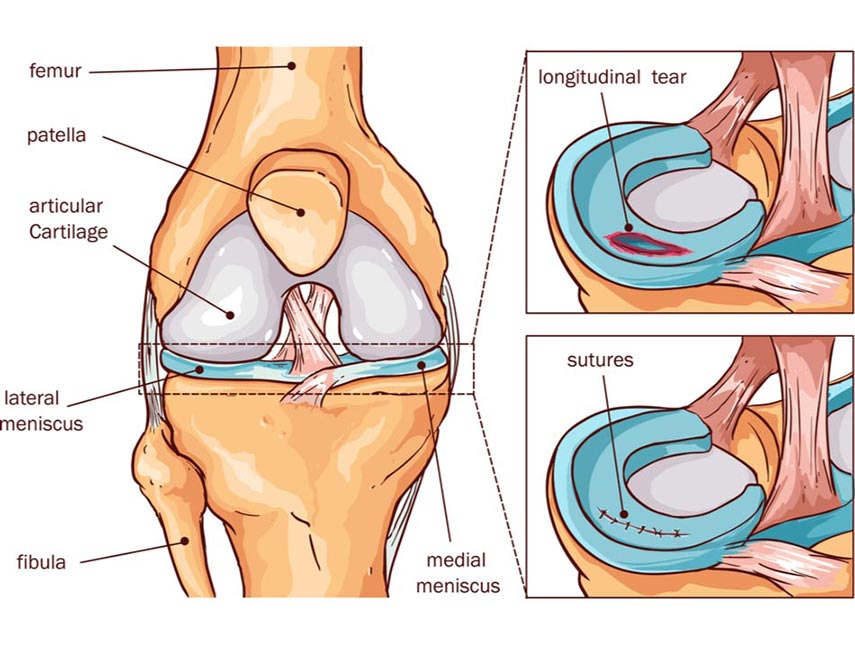
It is usually a relatively straightforward surgery that involves the damaged areas of the meniscus being repaired with stitches (most of the time); occasionally, the damaged area may have to be removed in more severe injuries. After the operation, patients are able to move their knees well, and many return to normal activities and sports.
Microfracture of osteochondral lesion
The bones in the knee (like most joints) are covered in hyaline cartilage, a type of smooth cartilage that allows for painless and smooth movement of your joints. Osteochondral lesions are localised injuries to the cartilage that can be caused by trauma or overuse of the knee, and may cause pain, swelling, and stiffness when you use the joint (for example when walking and climbing stairs).
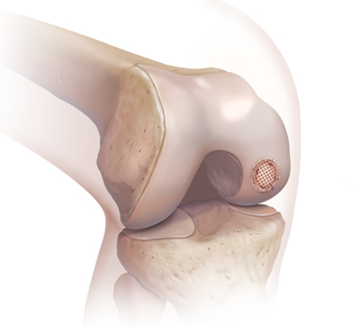
Your surgeon may offer a procedure called microfracture, a surgery done via a “keyhole” incision, which involves multiple 1-2mm small drill holes being created inside the knee bones, to allow stem cells from the bone to enter the joint. This stimulates regrowth of the cartilage over time and usually helps to restore pain-free movement & walking.
Microfracture of osteochondral lesion
Similar to the knee discussion above, arthroscopic microfracture surgery for ankle osteochondral lesions is a procedure that is helpful for patients with ankle joint pain stemming from localised cartilage damage due to trauma or overuse. After this surgery, you should be able to walk with less pain, have a better range of motion and benefit from restored function in your ankle joint!
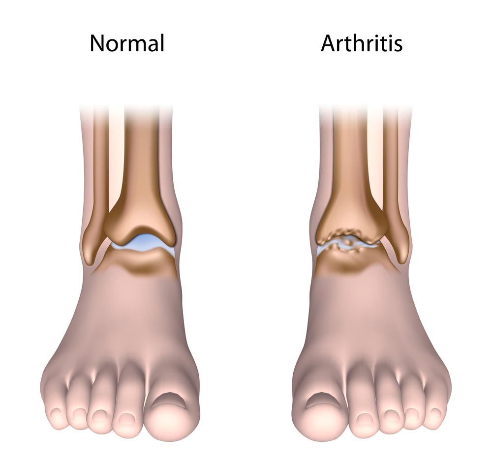
Fusion (also called arthrodesis) of the ankle joint involves the surgeon removing the damaged areas of cartilage and bone in the arthritic ankle joint, then fixing the bones of the ankle joint together (with screws and occasionally plates) to stimulate both bones to join together (fuse) into a single bone. This creates a stable ankle that removes the pain, and allows for reliable return to walking and most recreational sports!
An expected side effect of ankle fusion is a reduced range of motion of the ankle joint since the joint has been fused. However, the pain relief and restoration of ankle stability far outweigh the stiffness, and you will experience an improvement in walking and return to daily activities after the surgery.
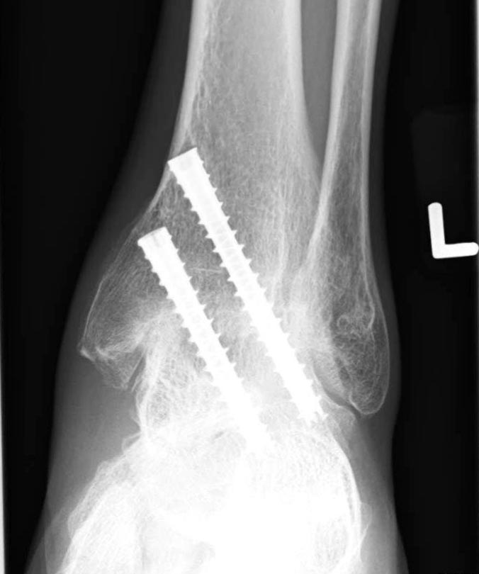
Ankle fusion (arthrodesis) is most commonly performed by conventional open surgery via a 10-15cm incision over the ankle. With modern technical advances, arthroscopic ankle fusion is becoming the new standard of care, as minimally invasive “keyhole” surgery markedly reduces the skin complications and infections that are problematic in open surgery. By only using several subcentimetre incisions, arthroscopic ankle fusion has the further benefits of less pain, more rapid recovery, earlier discharge home (often within a day), and better fusion rates compared to open ankle fusion.
Subtalar fusion, Talonavicular fusion & Calcaneocuboid fusion (Arthritis, Pes planus, Pes cavus)
Just like any other joint in the body, the foot is also susceptible to arthritis and damage to the cartilage over many years of strain and trauma. If severe, it can greatly affect your quality of life and limit your ability to walk properly.
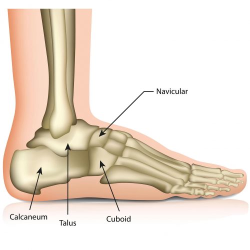
The talus is one of the important load-bearing foot bones and is involved in 2 important hind foot joints – 1) subtalar joint (the joint below the talus) is involved in the sideways tilting movement (inversion & eversion) of the foot; 2) talonavicular joint (the joint in front of the talus) is involved in complex tri-planar movements of the foot. Lastly, the calcaneocuboid joint lies adjacent to both the subtalar and talonavicular joints, and is also involved in tri-planar movements of the foot. One, two or all of these joints may be damaged in foot injuries and chronic conditions such as arthritis, pes planus (flat foot) and pes cavus (high-arched foot), and all can be treated effectively with arthroscopic surgery.
In subtalar, talonavicular and/or calcaneocuboid fusion surgery (also called triple fusion or triple arthrodesis), the areas of damaged bone & cartilage in these joints are carefully removed. After restoring the correct shape of the foot, the subtalar, talonavicular and/or calcaneocuboid joints are held in place with metal screws or plates. Similar to ankle fusion (described above), the bones in these joints will fuse together, resulting in greater stability and reliable pain relief.
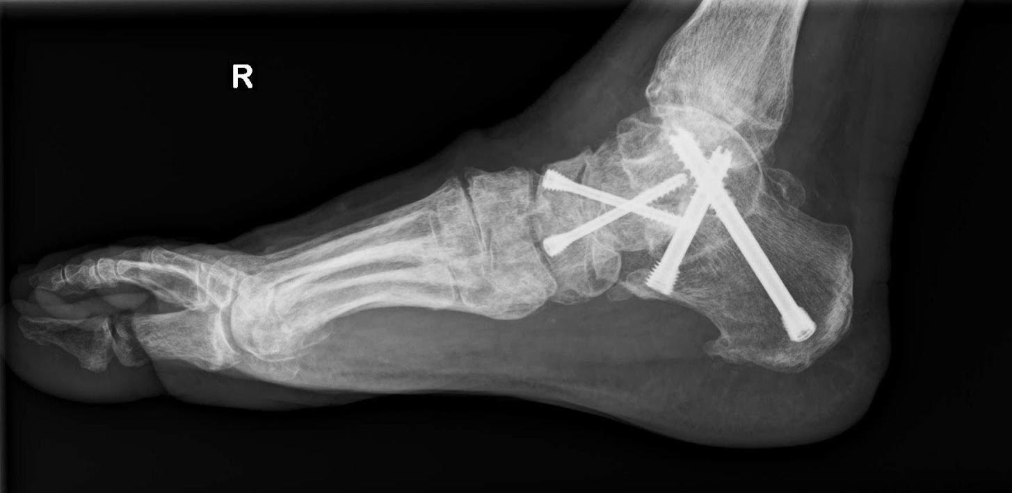
When done using the “keyhole” technique, arthroscopic triple fusion has much lower skin complications and infections that are problematic with open surgery. Arthroscopic triple arthrodesis has the additional benefits of much less pain, more rapid recovery, earlier discharge home (often within a day), and better fusion rates. Patients typically report better and faster return to activity and work following arthroscopic triple fusion.
Owing to the relatively small size and complex shape of the subtalar joint, calcaneocuboid joint, and particularly the talonavicular joint, arthroscopic triple fusion of these joints is very technically demanding surgery that requires significant expertise in foot arthroscopy, and hence is offered by only a very small number of foot & ankle surgeons locally. Please check with your foot & ankle surgeon if you are considering arthroscopic triple arthrodesis surgery.
If you have any of the conditions listed above or other joint injuries, do arrange for a consult with your doctor to discuss your treatment options and assess your suitability for this procedure.